html
Specialized in retinal surgery
All information on eye pathologies and treatments.
Red eye · Sudden loss of vision · Eye trauma · Double vision · Floaters or flashes
Hospitals of 15-20
One of the best ophthalmologists in Casablanca. Trained in leading Parisian establishments and certified by the European Council.
2018 – 2020 · Specialist assistant
2014 – 2018 · Intern — Internship specialized in Ophthalmology
2020 – 2022 · University clinical head-hospital assistant
2018 – 2020 · Specialist assistant — Professor Baudouin / Professor Labbé
Fellow of the European Board of Ophthalmology
DU Imaging · DU Glaucoma · IUD Refractive Surgery
Are you a demanding athlete and every detail counts to achieve excellence? Refractive surgery frees you from glasses and lenses for clear vision, without discomfort or distraction. No more worrying about fogging, sweating or uncomfortable lenses during exercise.
Every detail counts, and your eyesight is your first tool. Refractive surgery frees you from slipping glasses or uncomfortable lenses, for clear and precise vision, without discomfort, from morning to evening.
Give yourself the freedom of clear vision without glasses or lenses. Thanks to refractive surgery, reveal your eyes and gain daily comfort, without compromising on style.
Your daily life is more active than ever, but your glasses are holding you back? Free yourself from the constraints of glasses and lenses with refractive surgery. Clear vision, greater comfort, and the freedom to live every moment to the fullest.
Your job leaves no room for imprecision: every detail counts, every fraction of a second too. Thanks to refractive surgery, you regain clear, rapid and natural vision.
Tired of hiding your eyeliner behind glasses? With refractive surgery, enjoy clear vision without glasses, to dare all looks, enhance your eyes and let your creativity speak.
Myopia, hyperopia, astigmatism — all ametropias are treatable depending on your corneal profile.
MAKE AN APPOINTMENT FOR AN ASSESSMENTTrained at the Center Hospitalier National des 15-20 and at the Rothschild Foundation in Paris. FEBO (Fellow of the European Board of Ophthalmology) certified. Specialized in refractive surgery (LASIK, SMILE, PKR) and retinal surgery.
Dr Mehdi Batras · Cabinet Maarif, Casablanca · 07 11 10 50 07
Treatments and surgeries of the eye, retina and eyelids.
Interventions in the posterior segment of the eye (vitreous and/or retina) are currently carried out endoocularly using a technique called vitrectomy.
The procedure takes place on an outpatient basis in the clinic (during half a day), in the operating room under a microscope, and under regional anesthesia. In certain cases, the ophthalmologist in Casablanca Dr Batras can combine retinal surgery with cataract surgery.
Detachment, macular hole, membrane, AMD, diabetes
Cataract, myopia, hyperopia, astigmatism, refractive
Strabismus, glaucoma, eyelid surgery
OCT, ultrasound, lenses, fundus
HD Imaging
Fluorescein, ICG
Photocoagulation
Accuracy
If you are considering treatment for your eyes in Casablanca, here is some practical information.
Find all our articles and advice from Dr Mehdi Batras.
Discover the reviews on Google of patients of Dr Mehdi Batras ophthalmologist in Casablanca.
“Competent ophthalmologist, attentive, takes the time to explain and very patient with little ones. The office is clean and appointments are kept. »
“A big thank you for seeing me urgently at the office at the end of the day. I consulted the ophthalmologist Dr. Batras in his office in Casablanca following a retinal problem. Very attentive and professional, he took the time to explain my problem to me with great kindness. I was taken care of quickly, I recommend this ophthalmologist to everyone. Thanks also to his adorable assistant 🙏🏼. »
“My mother was lucky enough to have surgery by Dr. Batras for a serious retinal problem, and we couldn't be more grateful. Thanks to his expertise and dedication, my mother's vision was saved. The Doctor was exceptional from start to finish, always available to answer our questions and reassure us. We highly recommend this eye surgeon for his professionalism and humanity. Moroccans no longer need to go to Europe to carry out this type of operation! With Doctor Batras, Europe comes to your home to treat you ☺️ »
« I went with my husband and we were both very satisfied 👍🏻 with this ophthalmologist. »
Ophthalmologist in Casablanca
You can consult an ophthalmologist to:
There is no difference between an ophthalmologist and an ophthalmologist. These two terms refer to a doctor specializing in the diagnosis, treatment of eye diseases and correction of vision disorders.
An annual check-up at least once a year with an ophthalmologist is necessary to ensure good health of your eyes.
The ophthalmologist is a doctor specializing in the diagnosis and treatment of eye conditions, while the orthoptist is a paramedical health professional dedicated to visual rehabilitation and the prevention of vision disorders.
The office of Dr Mehdi Batras is considered one of the best ophthalmology offices in Casablanca, offering ophthalmology and orthoptics treatments.
Regular eye examination is essential to quickly detect possible disorders or degenerative diseases, such as AMD.
The eye examination carried out by the ophthalmologist consists of evaluating visual acuity, eye movements, visual fields, external structures of the eye and the retina. The fundus of the eye is inspected with an ophthalmoscope.
As the eye is the only transparent organ in the body, the examination can provide information on the patient's general health: detection of brain tumors, high blood pressure, bacterial infections, multiple sclerosis, even drug poisoning.
After the examinations, the ophthalmologist will be able to analyze the results and determine the most appropriate treatment.
Our team is at your disposal.
Ourika Residence 1st floor, 1 Corner Bir Anzarane and Abou Ghaleb Chiani, Maarif – Casablanca
Fixed: 05 20 70 55 66
Mobile: 07 11 10 50 07
Mon – Fri: 9 a.m. – 6 p.m. · Sat: 9 a.m. – 1 p.m.
Method of meeting:
07 11 10 50 07
Book online 24 hours a day
Fixed & Portable
[email protected]
Retinal detachment is a rare but serious and potentially blinding condition characterized by the accumulation of fluid under the retina.
Retinal detachment is a rare ocular pathology, characterized by the separation of the retina from the outer membranes of the eyeball. This break prevents the retina from functioning, leading to vision problems. If not treated quickly, it can progress to permanent blindness.
Vitrectomy takes place under local or general anesthesia, on an outpatient basis or with a short hospitalization. The surgeon begins by removing the vitreous (transparent gel that fills the eye), which allows free access to the detached retina. The tears responsible for the detachment are then treated by laser photocoagulation or cryotherapy to create a scar which will reattach the retina. A bubble of expanding gas or silicone oil is then injected to press the retina against the wall of the eye during healing. The procedure lasts on average between 1 hour and 2 hours depending on the complexity of the detachment.
A specific head position (often face down or sideways) must be maintained for several days for the gas bubble to press effectively against the retina. Vision remains blurry and distorted for several weeks while the gas resolves and the retina heals. Antibiotic and anti-inflammatory eye drops are prescribed for about a month. Air travel is strictly prohibited while the gas is present in the eye. The final visual recovery lasts 3 to 6 months and essentially depends on the age of the detachment and the involvement of the macula.
Here is the essential thing to know about diabetes and its effects on eyesight.
Diabetes is a chronic disease characterized by high blood glucose levels due to insulin dysfunction. There are two types: type 1 (pancreas does not produce insulin) and type 2 (insufficient or incorrectly used insulin). Diabetes can cause cardiovascular, nervous and vision problems.
Type 1: insulin by injection or pump. Type 2: oral medications, dietary changes, exercise. Regular blood sugar monitoring essential. Continuous medical monitoring to prevent complications.
Ophthalmologist Casablanca specializing in retina
Mon–Fri 9 a.m.–6 p.m. · Sat 9 a.m.–1 p.m. · Maarif, Casablanca 20100
Make an appointmentSmall (< 150 µm) or large, this pathology must be treated quickly enough to close the hole. Find out how our firm can help you.
The macular hole is a small tear or opening that forms in the macula, the central region of the retina responsible for fine, detailed vision. Causes may include aging, eye trauma, or retinal diseases. There are several treatments to repair the hole and improve vision.
These symptoms sometimes progress slowly.
Treatment is generally done by vitrectomy. After surgery, the patient is often required to maintain a specific position, and eye drops are prescribed to promote healing.
The procedure lasts approximately 30 to 45 minutes under local anesthesia, on an outpatient basis. The surgeon first performs a 3-way vitrectomy using micro-incisions of less than 1 mm to remove the vitreous. He then very gently peels back the internal limiting membrane around the hole using microsurgical forceps — a key step that requires extreme precision. A bubble of expansive gas is finally injected into the vitreous cavity to buffer the hole from the inside and promote its spontaneous closure. No sutures are necessary thanks to the fineness of the incisions.
The face down position is essential for 5 to 10 days so that the gas bubble presses on the hole and promotes its closure — this is the key to success. Vision remains blurry for several weeks, then gradually improves over 2 to 3 months. Air travel is prohibited as long as the gas is present. Eye drops are prescribed for about a month, and an OCT check makes it possible to check that the hole is closed correctly.
Ophthalmologist Casablanca specializing in retina
Mon–Fri 9 a.m.–6 p.m. · Sat 9 a.m.–1 p.m. · Maarif, Casablanca 20100
Make an appointmentA common benign condition of the retina, often associated with aging. Find out how we can help you.
The epiretinal membrane is a thin layer of fibrocellular tissue that forms on the surface of the retina, more precisely on the macula, the area responsible for central vision. This condition, usually related to age, can also result from inflammation, previous eye surgery or retinal detachment.
Symptoms often appear gradually:
Carried out on an outpatient basis under local or regional anesthesia, the procedure lasts 30 to 45 minutes. The surgeon first removes the vitreous by 3-way vitrectomy, then gently peels the fibrous membrane from the surface of the retina using microsurgical forceps under a microscope. This step requires great precision so as not to damage the underlying retina. Depending on the case, the internal limiting membrane can also be peeled to reduce the risk of recurrence. No gas tamponade is generally necessary at the end of the procedure.
No particular position is imposed after the intervention. Vision remains blurry and distorted in the first weeks, then gradually improves over 3 to 12 months depending on the age of the membrane and the initial state of the macula. Anti-inflammatory and antibiotic eye drops are prescribed for about a month. Regular OCT checks make it possible to monitor anatomical and functional recovery. Recovery of visual acuity can be gradual but lasting.
Ophthalmologist Casablanca specializing in retina
Mon–Fri 9 a.m.–6 p.m. · Sat 9 a.m.–1 p.m. · Maarif, Casablanca 20100
Make an appointmentAge-related macular degeneration (AMD) is the leading cause of low vision in people over 50.
AMD is an eye condition that affects the macula, the central part of the retina responsible for fine, detailed vision. It causes a progressive loss of central vision. Generally linked to aging, genetic and environmental factors also play a role. There are two forms: dry AMD (more common) and wet AMD (more serious, rapid loss of vision).
Regular follow-up with the doctor is essential to monitor progress and adjust treatment.
For exudative (wet) AMD, treatment is based on intravitreal injections of anti-VEGF which block the growth of abnormal new vessels. The injection is carried out in the office in a few minutes, under topical anesthesia in sterile conditions. A thin needle delivers the medication directly into the vitreous. The molecules used (Eylea®, Lucentis®, Beovu®) are chosen according to the patient's profile. The injections are first close together (3 consecutive months) then gradually spaced out depending on the response, on average every 4 to 8 weeks to stabilize the disease in the long term.
A slight redness, discomfort or blurred vision is normal for 24 to 48 hours after the injection — it is not serious. The effectiveness of the treatment is monitored by monthly or bimonthly macular OCT to adapt the rhythm of the injections as accurately as possible. The objective is to stabilize vision, or even improve it. Follow-up is lifelong because AMD is a chronic disease. Self-monitoring with an Amsler grid makes it possible to quickly detect any evolving recovery between checks.
Ophthalmologist Casablanca specializing in retina
Mon–Fri 9 a.m.–6 p.m. · Sat 9 a.m.–1 p.m. · Maarif, Casablanca 20100
Make an appointmentRefractive surgery aims to make patients independent of glasses or contact lenses.
Refractive surgery includes interventions aimed at correcting refractive disorders, using devices such as lasers or implants, as well as surgical instruments.
All ametropia (visual defects) can currently be corrected by refractive surgery. The patient must be at least 21 years old and have a visual defect (myopia, hyperopia, astigmatism) that has been stable for at least 2 years. Additional examinations eliminate contraindications.
The surgeon carries out a careful history and clinical examination to eliminate any associated pathology. Corneal examinations are carried out: corneal pachymetry (thickness measurement) and corneal topography to rule out keratoconus. Wearing lenses must be stopped several days before the assessment.
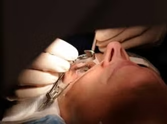
The procedure takes place on an outpatient basis, under local anesthesia (drops). Both eyes are operated on at the same time.
Ophthalmologist Casablanca specializing in retina
Mon–Fri 9 a.m.–6 p.m. · Sat 9 a.m.–1 p.m. · Maarif, Casablanca 20100
Make an appointmentDiscover the solutions that ophthalmologist Dr Batras offers for this eye disease which affects one in 5 people after the age of 65.
Cataract is one of the main causes of vision loss. It is characterized by a progressive opacification of the crystalline lens, the natural lens of the eye. This phenomenon can occur naturally with age. Although often linked to aging, it can also develop for other reasons: congenital cataracts, diseases such as uveitis or diabetes, corticosteroids, or eye trauma.
It evolves gradually:
Thanks to precise examinations, Dr. Batras can diagnose an incipient cataract in each patient.
Surgery is mainly indicated. The diagnosis is clinical by biomicroscopy, with pupil dilation to examine the lens and the fundus. Photographs and OCT can be performed to rule out other pathologies.
Phacoemulsification lasts approximately 15 to 20 minutes under topical anesthesia (anesthetic eye drops), on an outpatient basis and without hospitalization. The surgeon makes a micro-incision of only 2.2 mm, then fragments the clouded lens using ultrasound and aspirates it in a few minutes. A foldable intraocular implant is then placed - its type is chosen according to your vision and your needs (monofocal, multifocal, toric for astigmatism). The incision is so fine that it closes spontaneously without suturing. The procedure is completely painless.
Mild discomfort and sensitivity to light are normal for the first day. Vision generally improves the next day or within 48 hours, and continues to stabilize over 2 to 4 weeks. Antibiotic and anti-inflammatory eye drops are prescribed for 3 to 4 weeks. It is important to avoid rubbing the eye, swimming in the sea or swimming pool and violent physical exertion for 3 to 4 weeks. A protective shell is worn at night for the first week. Permanent glasses are prescribed approximately one month after the procedure if necessary.
Ophthalmologist Casablanca specializing in retina
Mon–Fri 9 a.m.–6 p.m. · Sat 9 a.m.–1 p.m. · Maarif, Casablanca 20100
Make an appointmentThis is one of the most common pathologies in ophthalmology. Find out how we can help you.
Myopia is a vision disorder where nearby objects are seen clearly, but distant objects appear blurry. This happens when the eyeball is too long or the cornea is too curved, causing light rays to focus in front of the retina rather than directly on it.
These symptoms may appear gradually and get worse over time.
Myopia is mainly treated with glasses or contact lenses. For more permanent correction, refractive surgery such as LASIK or PRK may be considered, reshaping the cornea to improve vision.
Ophthalmologist Casablanca specializing in retina
Mon–Fri 9 a.m.–6 p.m. · Sat 9 a.m.–1 p.m. · Maarif, Casablanca 20100
Make an appointmentDiscover the solutions we offer to help you see more clearly.
Hyperopia is a vision disorder where nearby objects appear blurry while distant objects are seen more clearly. This happens when the eyeball is too short or the cornea is insufficiently curved, causing light rays to focus behind the retina.
These symptoms become more apparent during tasks requiring prolonged close vision.
Hyperopia is treated with glasses or convex contact lenses. For a permanent solution, procedures like LASIK or PRK may be considered, reshaping the cornea to improve near vision.
Ophthalmologist Casablanca specializing in retina
Mon–Fri 9 a.m.–6 p.m. · Sat 9 a.m.–1 p.m. · Maarif, Casablanca 20100
Make an appointmentAstigmatism is a very common visual defect that causes blurred or distorted vision at all distances. Discover our solutions.
Astigmatism is a refractive disorder caused by an irregularly shaped cornea (oval rather than round). Instead of focusing light into a single point on the retina, the astigmatic eye creates multiple points of focus, resulting in a blurry or distorted image. It can be associated with myopia or hyperopia.
These symptoms can be present from childhood and gradually worsen.
Ophthalmologist Casablanca specializing in retina
Mon–Fri 9 a.m.–6 p.m. · Sat 9 a.m.–1 p.m. · Maarif, Casablanca 20100
Make an appointmentA disorder where the visual axes of the eyes are not parallel. Discover the solutions we offer you.
Strabismus is a disorder where the eyes are not aligned correctly, not pointing in the same direction. This can lead to problems with visual coordination, impaired depth perception, and difficulty with binocular vision. It can affect one or both eyes and manifest as one eye deviating inwards, outwards, up or down.
Regular follow-up with an ophthalmologist is essential to assess progress and adapt treatment.
Ophthalmologist Casablanca specializing in retina
Mon–Fri 9 a.m.–6 p.m. · Sat 9 a.m.–1 p.m. · Maarif, Casablanca 20100
Make an appointmentGlaucoma is a chronic disease responsible for potentially blinding optic neuropathy. The main challenge lies in its detection and early treatment.
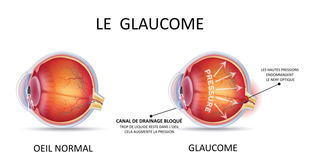
Glaucoma is a chronic and progressive optic nerve disease causing irreversible damage to the visual field. The main risk factor is ocular hypertonia (raised intraocular pressure). The most common is primary chronic open-angle glaucoma. There are also normal pressure, angle closure, and secondary glaucomas.
Glaucoma is often called the “silent thief of sight” because it can progress without obvious symptoms. Warning signs:
Who is affected? Anyone can be affected. Risk factors: ocular hypertonia, family history, age 55–75 years, high myopia, thin corneas, sleep apnea. Screening recommended after age 40.
Examinations: intraocular pressure (IOP), pachymetry, gonioscopy, fundus, retinophotography, OCT of the optic nerve and visual field.
Treatments: Glaucoma cannot be cured but is stabilized:
Ophthalmologist Casablanca specializing in retina
Mon–Fri 9 a.m.–6 p.m. · Sat 9 a.m.–1 p.m. · Maarif, Casablanca 20100
Make an appointmentThe eyelids can be affected by various disorders, ranging from tumor pathologies to malposition problems. Discover the solutions.
The eyelids are mobile structures that cover and protect the eyes. They regulate light entering the eye and distribute tears to maintain hydration and remove debris. They are made up of skin, muscles and glands.
Ophthalmologist Casablanca specializing in retina
Mon–Fri 9 a.m.–6 p.m. · Sat 9 a.m.–1 p.m. · Maarif, Casablanca 20100
Make an appointment
The international diabetes community is paying increasing attention to the psychosocial impacts of chronic diseases.
These impacts are partly related to physical manifestations and fluctuations in glycemic control, but also to individual factors, including the person's ability to manage both technically and emotionally the mental load associated with diabetes.
This article explores the notion of “diabetes-related distress”, ways to recognize it, its prevalence in people with type 1 diabetes, as well as approaches to its management.
Diabetes-related distress refers to all the negative emotions and psychological burden felt by people living with diabetes. It is not a psychiatric disorder in itself, but reflects a normal reaction to the daily demands of managing the illness.
Treatment is based on a multidisciplinary approach combining:
It is essential that health professionals systematically screen for this distress during consultations, because it directly impacts glycemic control and quality of life.
Dr Mehdi Batras — Ophthalmologist in Casablanca specializing in retinal surgery. For any questions about diabetes and its effects on eyesight, contact our office.
Machine learning is revolutionizing preoperative evaluation in refractive surgery.
Machine learning is a general technique for finding suitable parameters or functions to classify input data from large amounts of training data.
Several methodologies have been recommended:
Deep learning deals with the training of multi-layer artificial neural networks. Convolutional neural networks have achieved impressive results in image classification in ophthalmology and many scientific fields.
In refractive surgery, these technologies allow:
Dr Mehdi Batras — Ophthalmologist in Casablanca, refractive and retina surgery. Contact our office.
The rapid progression of myopia in children and adolescents constitutes a global public health issue. By 2050, nearly 50% of the world's population could be myopic. Several therapeutic strategies now make it possible to slow its progression.
The increase in the prevalence of myopia is closely linked to the increase in near vision time, increased use of screens and reduced exposure to daylight. With a projection of 1 billion highly myopic people in 2050, at high risk of potentially blinding complications, the objective of braking takes on its full meaning.
Early recognition of myopia allows treatment and prevention measures to be put in place. Parents must be particularly vigilant when they themselves are myopic. The initial examination should include: subcyclopegic refraction, intraocular pressure, axial length and fundus. Follow-up every 6 months is recommended.
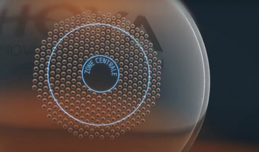
DIMS (Miyosmart® – Hoya) and HAL (Stellest® – Essilor) defocusing lenses: these lenses induce peripheral myopic defocus to slow down axial elongation. The study by Lam et al. (2020) shows a 52% reduction in myopic progression and 62% in axial elongation. Miyosmart® glass recently received an ASA IV score from the ANSM. Level of evidence: high (GRADE: High).
Defocusing lenses (MiSight®, Abiliti®, Bloom Oneday®): they slow down myopia by retinal defocus. The trial by Chamberlain et al. (2019) over 3 years shows a 59% reduction in progression. Level of evidence: high.
Wearing rigid lenses at night temporarily reshapes the cornea. The Cochrane meta-analysis (2023) concludes that there is a significant reduction in axial elongation: –0.19 mm at 1 year, –0.28 mm at 2 years. Level of evidence: moderate to high. Beware of the risk of infection among adolescents who are not compliant with hygiene.
Topical atropine at low doses (0.01% to 0.05%) acts by modulating scleral growth. LAMP studies demonstrate maximum effectiveness at 0.05%. 5-year follow-up indicates a stable safety profile. The APPLE study is the first to demonstrate effectiveness at lower concentrations: 0.01% and 0.005%. Level of evidence: high.
Faced with myopia that remains progressive under mono-treatment, combined therapies (defocusing lenses + atropine, lenses + atropine, orthokeratology + atropine) provide superior results. This integrated approach could become the norm.
Current recommendations from the WSPOS and the International Myopia Institute emphasize individualized care integrating epidemiological data, risk factors and intervention possibilities. The treatment decision is made in agreement with the parents and the child.
Dr Mehdi Batras — Ophthalmologist in Casablanca. For a myopia assessment of your child or to discuss braking options, contact our office.
Absolute emergency: if you suddenly lose vision in one eye, call SAMU (15) or go to the emergency room within 4.5 hours. Every minute counts.
Central retinal artery occlusion (CRAO) is often called the “stroke of the eye.” It is a rare but serious medical emergency, which can lead to irreversible vision loss if not treated immediately.
The retina is supplied by the central retinal artery. When this artery becomes blocked – most often by a clot (embolism) coming from the heart or carotids – the retina is no longer supplied with oxygen. Within minutes, retinal cells suffer and begin to die. Without prompt treatment, vision loss may be permanent.
CRAO shares the same mechanisms as cerebral stroke: this is why it must be managed as a neurovascular emergency. The risk of having a stroke in the 2 months following an OACR is multiplied by 9.
Illustration: occlusion of the central superior retinal artery with non-perfused area
Warning: loss of vision is often painless. The absence of pain can lead people to believe wrongly that it is not serious. This is a dangerous mistake.
An OACR sometimes repermeabilizes in 3 to 4 weeks. Regular ophthalmological follow-up is essential to monitor progress and prevent complications such as neovascularization. Your doctor will adapt the treatment according to the cause identified.
Dr Mehdi Batras — Ophthalmologist in Casablanca, specialized in retinal surgery. For any sudden eye symptoms, our office is available urgently. Contact us.
Mon–Fri 9 a.m.–6 p.m. · Sat 9 a.m.–1 p.m. · Emergencies: 07 11 10 50 07
Make an appointmentOCT is a non-invasive and painless imaging test that provides high-resolution sections of the retina, the optic nerve and the anterior segment of the eye, in a few seconds and without contact.
OCT makes it possible to detect and monitor numerous pathologies: AMD, diabetic macular edema, macular hole, epiretinal membrane, glaucoma (analysis of the nerve fiber layer) and retinal detachment.
You simply place your chin on a device, without contact with the eye, without injection or compulsory dilation. The exam lasts a few minutes and is completely painless.
Spectral domain OCT offers a resolution of the order of a micron, allowing early diagnosis and very precise monitoring of progress under treatment.
Ocular ultrasound uses ultrasound to visualize the internal structures of the eye, particularly when the ocular media (cornea, lens, vitreous) are not sufficiently transparent for direct examination of the fundus.
This examination is essential in cases of dense cataract, vitreous hemorrhage, suspicion of non-visible retinal detachment, or to measure the axial length of the eye before cataract surgery.
An ultrasound probe is gently applied to the closed eyelid (mode B) or directly to the eye after topical anesthesia (mode A). The exam is quick and painless.
Ultrasound allows you to “see through” an opaque eye and detect lesions, masses or separations that would not otherwise be visible.
The adaptation of contact lenses is made to measure according to the morphology of your eye, your correction and your lifestyle, for optimal comfort and clear vision all day long.
Lenses correct myopia, hyperopia, astigmatism and presbyopia. They are also used to reduce myopia in children (defocusing lenses such as MiSight®).
Corneal topography and precise refraction allow you to choose the appropriate type of lens (soft, rigid, daily, monthly). A test is carried out in the office with tolerance control.
Regular check-ups are essential to prevent any risk of corneal infection, especially among wearers of soft or orthokeratology lenses.
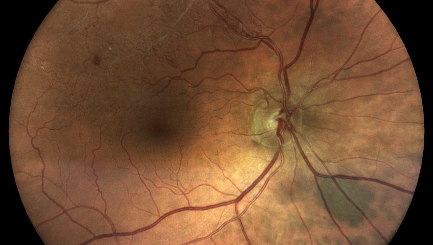
The fundus examination consists of dilating the pupil using specific drops to observe, as its name suggests, the fundus of the eye: retina, macula, vessels and optic nerve.
The fundus examination helps detect diabetic retinopathy, AMD, glaucoma, retinal tears and detachments, as well as signs of high blood pressure or vascular diseases.
Dilating drops are instilled 15 to 20 minutes before the examination. Vision will be blurry and the light will be dazzling for a few hours – if possible, plan not to drive immediately afterwards.
Annual fundus examination is recommended in patients with diabetes, hypertension, high myopia or a family history of glaucoma or AMD.
OCT (Optical Coherence Tomography) Spectral Domain is a cutting-edge imaging technology allowing high-resolution sections of the retina and optic nerve to be obtained, in a few seconds and without any contact with the eye.
Spectral OCT uses an infrared light beam to scan the retina and reconstruct cross-sectional images (like a very high resolution optical “ultrasound”, in the order of a micron). Unlike traditional ultrasound, OCT does not use ultrasound but light, which allows incomparable precision at the cellular level.
OCT is today essential for the diagnosis and monitoring of numerous pathologies: AMD (dry and exudative), diabetic macular edema, epiretinal membrane, macular hole, retinal vascular occlusions, glaucoma (analysis of the optic nerve fiber layer), retinal detachment and central serous chorioretinopathy.
The examination is completely painless and contactless. You rest your chin on a chin rest and stare at a lighted target while the device automatically scans your retina. Pupil dilation is usually not necessary. The examination lasts 5 to 10 minutes on both eyes, and the results can be immediately analyzed by your ophthalmologist.
Angiography is an examination which allows the blood circulation in the retina and choroid to be visualized using the intravenous injection of a fluorescent dye (fluorescein or indocyanine green).
A fluorescent contrast product is injected into a vein in the arm, then circulates rapidly throughout the body and reaches the retinal vessels within a few seconds. A special camera then takes a series of images in blue (fluorescein) or infrared (indocyanine green) light to visualize blood flow and detect possible vascular abnormalities.
Angiography is essential to explore diabetic retinopathy, exudative AMD (search for new vessels), retinal venous or arterial occlusions, retinal vasculitis, certain intraocular tumors, and central serous chorioretinopathy. It is often an essential complement to OCT when deciding on laser or injection treatment.
Pupil dilation is necessary (drops 15 to 20 minutes before). The contrast product is then injected over a few seconds, then images are taken continuously for 5 to 10 minutes. The examination is painless but can cause a temporary yellowish discoloration of the skin and urine (24 to 48 hours) with fluorescein. A slight sensation of warmth may be felt at the time of injection.
The multispot laser is a new generation retinal photocoagulation technology which makes it possible to deliver several laser impacts simultaneously, in a very short time and with optimal comfort for the patient.
Unlike the conventional laser which delivers a single impact at a time, the multispot laser can project a grid of 4 to 25 spots in a few milliseconds. Shorter pulse durations (10 to 30 ms) significantly reduce thermal diffusion to surrounding tissues, which improves comfort, reduces pain and better preserves the healthy retina.
The multispot laser is used for panretinal photocoagulation (PPR) in proliferative diabetic retinopathy, retinal tears (laser barrier), ischemic venous occlusions, and certain peripheral lesions at risk of detachment. It has become the benchmark for treating large retinal surfaces in a single session.
The procedure takes place in consultation, under topical anesthesia (anesthetic eye drops) and pupil dilation. The patient sits facing the laser with his chin rested. A special contact lens is applied to the eye to focus the beam. The session lasts 10 to 20 minutes depending on the extent to be treated. A slight discomfort or light sensation is normal during and after treatment, but fades quickly.
The operating microscope is the central surgical tool for any ophthalmological operation. The latest generation models offer high-resolution 3D visualization, adaptive lighting and the integration of real-time intraoperative imaging.
The modern operating microscope allows up to 20 times magnification with optimal depth of field and sharpness. It incorporates LED light sources with adjustable intensity, filters to reduce retinal phototoxicity, and can be coupled with intraoperative OCT ("integrated OCT") which allows the surgeon to visualize the layers of the retina live during the procedure.
The operating microscope is essential for all ophthalmological surgeries: cataract (phacoemulsification), retinal surgery (vitrectomy, retinal detachment, macular hole, epiretinal membrane), glaucoma surgery, corneal transplantation, and refractive surgery. The quality of the microscope directly determines the precision of the surgical procedure and therefore the safety of the procedure.
The microscope is positioned above the patient and the surgeon operates seated, looking through the eyepieces (or a 3D screen depending on the model). Foot control integration allows you to zoom, focus, pan and adjust lighting without using your hands. This technology allows for more precise, safer and faster surgery than with previous generation microscopes.
Hospital specialist assistant — Professor Baudouin
The Quinze-Vingts National Ophthalmology Hospital Center is the benchmark establishment in ophthalmology in France and one of the most prestigious in the world. Founded in 1260 by Saint Louis, it is today the national reference center for complex and rare ocular pathologies.
As an assistant specialist under the direction of Professor Baudouin, Dr. Batras completed his surgical training there in the fields of cataract, medical and surgical retina, glaucoma and refractive surgery. The department treats hundreds of patients per day and carries out a considerable surgical volume.
Intern — Internship specialized in Ophthalmology
The Rothschild Foundation is a private health establishment of collective interest, one of the largest ophthalmology centers in Europe. Renowned for its clinical and scientific excellence, it performs more than 40,000 surgical procedures per year, in all areas of ophthalmology.
Dr Batras completed his entire ophthalmology internship there, i.e. 4 years of intensive training. This time at the Rothschild Foundation formed the basis of his surgical training, with exposure to all subspecialties: cataract, medical and surgical retina, glaucoma, ophthalmology, refractive surgery and oculoplasty.
University clinical head-hospital assistant
The Simone Veil Hospital in Eaubonne (Nord Val d'Oise Hospital Group) is a hospital center attached to Paris-Sorbonne University, welcoming a large population in Île-de-France. Its ophthalmology department is recognized for its surgical activities and its university teaching.
As Head of University Clinic, Dr. Batras combined three missions: clinical and surgical care of patients, teaching of interns and medical students, and clinical research. This position allowed him to deepen his surgical expertise while passing on his knowledge.
Hospital specialist assistant — Professor Baudouin / Professor Labbé
The Ambroise-Paré Hospital, located in Boulogne-Billancourt, is part of the Assistance Publique–Hôpitaux de Paris (AP-HP). Its ophthalmology department is headed by professors Christophe Baudouin and Antoine Labbé, international references in the treatment of glaucoma, the ocular surface and the anterior segment.
Under the direction of Professors Baudouin and Labbé, Dr Batras perfected his mastery of the diagnosis and treatment of glaucoma (medical, laser and surgical), as well as pathologies of the ocular surface (dryness, blepharitis, corneal ulcers). This dual expertise enriches his daily practice at the office.
Fellow of the European Board of Ophthalmology
The FEBO (Fellow of the European Board of Ophthalmology) title is a European certification awarded by the European Board of Ophthalmology. It validates the theoretical and practical skills of an ophthalmologist according to criteria common to all of Europe.
To obtain the FEBO, the candidate must pass a rigorous examination including a written test (multiple choice questions and clinical cases) and an oral test before a jury of European experts. This certification attests to a level of knowledge aligned with the highest European standards.
The FEBO is recognized in all European countries and allows you to practice in an international framework. For patients, it is the assurance of being treated by a doctor trained according to the best quality standards and continually up to date with the latest scientific advances.
Three complementary specializations obtained in parallel with internship and hospital training, demonstrating in-depth expertise in specific areas of ophthalmology.
This university diploma trains in the expert interpretation of imaging examinations in ophthalmology: OCT (Optical Coherence Tomography), fluorescein and indocyanine green angiography, ocular ultrasound, OCT-angiography. It is essential for the diagnosis and monitoring of retinal, macular and optic nerve pathologies.
This specialized training delves into the management of glaucoma — a disease that progressively damages the optic nerve and can lead to blindness. The DU covers the entire discipline: screening, diagnosis, medical treatment (eye drops), laser treatments (SLT, iridotomy) and surgical treatments (trabeculectomy, deep sclerectomy, drainage valves).
This inter-university diploma trains in modern surgical techniques for correcting visual defects: LASIK, SMILE, PKR, but also phakic intraocular implants and clear lens surgery. It covers candidate selection, surgical planning, operative procedures and management of complications.
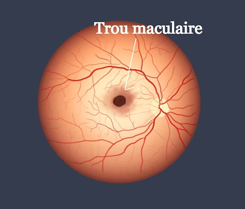
The macula is the central area of the retina, responsible for fine vision and details (reading, faces, driving). A macular hole is a small opening that forms in this area and distorts or blurs central vision. Although impressive, today it is a pathology that is very well managed surgically, with high success rates.
With age, the vitreous (the transparent gel that fills the inside of the eye) gradually shrinks and detaches from the retina. In most cases, this detachment occurs without problem. But sometimes, the vitreous remains stuck to the macula and pulls on it, until it creates a real opening. This is the most common cause (we speak of an “idiopathic” hole). More rarely, a hole may appear following trauma, high myopia, or in association with another retinal disease.
The macular hole manifests itself by a progressive decline in central vision, straight lines that appear wavy or broken (metamorphopsia), and sometimes a dark or blurry spot in the center of the eye. Peripheral vision remains normal – it is the center of the image that is affected. These symptoms warrant rapid consultation: the sooner the hole is treated, the better the chances of visual recovery.
The diagnosis is based on fundus examination, confirmed and clarified by macular OCT (optical coherence tomography). This painless and rapid examination makes it possible to precisely measure the size of the hole — an essential criterion, because the diameter directly influences the surgical strategy and the chances of success. We distinguish in particular between small holes (less than 400 microns) and larger holes, which sometimes require additional surgical techniques.
The standard treatment is vitrectomy: the surgeon removes the vitreous responsible for the traction, then delicately prepares the edges of the hole to promote its closure. A thin natural membrane present on the surface of the retina (the internal limiting membrane) is most often removed around its perimeter, which significantly increases the chances of success. The procedure ends with the injection of a gas bubble which “buffers” the hole from the inside, while it heals.
Certain holes are more difficult to close: large holes, recurrent holes after a first surgery, or those associated with severe myopia. In these situations, additional surgical techniques exist (use of biological grafts, retinal mobilization techniques) and today make it possible to obtain very good closure rates, even in cases previously considered difficult.
In the days following the operation, it is often asked to keep the head positioned downward for several days: this allows the gas bubble to press firmly on the hole and promote its definitive closure. Air travel should be avoided as long as the gas is present in the eye. Vision remains blurry for several weeks while the gas naturally resolves, then gradually improves over several months.
To remember: the earlier a macular hole is diagnosed and operated on, the better the visual prognosis. If you notice a distortion of straight lines or a decrease in central vision, consult quickly.
Glaucoma is a chronic eye disease that gradually damages the optic nerve, most often due to excessively high intraocular pressure. It is a silent disease: it causes no pain or symptoms at first, but can lead to irreversible vision loss if not treated in time. The good news: with appropriate treatment and regular monitoring, we can stop its progression.
In a healthy eye, a fluid (the aqueous humor) is produced and then evacuated constantly, maintaining a stable pressure. In glaucoma, this fluid is poorly evacuated: the pressure rises and compresses the optic nerve, which transmits images to the brain. Damaged nerve fibers do not regenerate — hence the importance of treating early. The goal of treatment is simple: to lower eye pressure to protect the optic nerve and preserve remaining vision.
Treatment almost always begins with eye drops to be instilled every day. Several families exist:
When a single eye drop is not enough, several can be combined, often in the form of a combined bottle to simplify treatment.
Glaucoma can be treated every day, for life. As the disease does not hurt, we are sometimes tempted to forget or stop our drops - this is the main cause of aggravation. Instilling your eye drops at a set time, closing your eyes 1 to 2 minutes after instilling (this doubles the effectiveness) and scrupulously following the prescription are essential to lastingly protect your eyesight.
The SLT laser (selective trabeculoplasty) is an alternative or complement to eye drops. In a few minutes, painless and in consultation, it improves the natural evacuation of liquid. It is particularly interesting when the drops are poorly tolerated or difficult to take regularly. Its effect lasts several years and the gesture can be repeated.
When eye drops and laser are no longer sufficient to control the pressure, or in cases of advanced glaucoma, surgery may be offered (trabeculectomy, deep sclerectomy, or installation of drainage devices). These interventions create a new evacuation route for the liquid and allow the pressure to be lowered sustainably.
Glaucoma treatment is personalized: it depends on the type of glaucoma, the level of pressure, the age and the tolerance of each patient. Regular monitoring (measurement of pressure, visual field, OCT of the optic nerve) makes it possible to check that the disease is well stabilized and to adjust the treatment if necessary. Detected and treated in time, glaucoma does not prevent you from maintaining good vision throughout your life.
Dr Mehdi Batras — Ophthalmologist in Casablanca, holder of a DU in Glaucoma. Regular screening after age 40 is recommended, especially if there is a family history. Make an appointment for an assessment.