Ophthalmology guide

Macular hole: understanding and treating

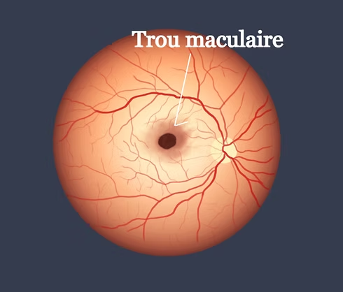
The macula is the central area of ​​the retina, responsible for fine vision and details (reading, faces, driving). A macular hole is a small opening that forms in this area and distorts or blurs central vision. Although impressive, today it is a pathology that is very well managed surgically, with high success rates.

Verified medical informationDr Mehdi Batras · Casablanca
Le trou maculaire : comprendre et traiter

Macular hole: understanding and treating

MB
Dr Mehdi Batras — Ophthalmologist Casablanca
August 2026 · 8 min reading · Patient article

UnderstandWhy does a hole form?

With age, the vitreous (the transparent gel that fills the inside of the eye) gradually shrinks and detaches from the retina. In most cases, this detachment occurs without problem. But sometimes, the vitreous remains stuck to the macula and pulls on it, until it creates a real opening. This is the most common cause (we speak of an “idiopathic” hole). More rarely, a hole may appear following trauma, high myopia, or in association with another retinal disease.

SymptomsHow to recognize it?

The macular hole manifests itself by a progressive decline in central vision, straight lines that appear wavy or broken (metamorphopsia), and sometimes a dark or blurry spot in the center of the eye. Peripheral vision remains normal – it is the center of the image that is affected. These symptoms warrant rapid consultation: the sooner the hole is treated, the better the chances of visual recovery.

DiagnosisOCT, the key examination

The diagnosis is based on fundus examination, confirmed and clarified by macular OCT (optical coherence tomography). This painless and rapid examination makes it possible to precisely measure the size of the hole — an essential criterion, because the diameter directly influences the surgical strategy and the chances of success. We distinguish in particular between small holes (less than 400 microns) and larger holes, which sometimes require additional surgical techniques.

TreatmentSurgery: vitrectomy

The standard treatment is vitrectomy: the surgeon removes the vitreous responsible for the traction, then delicately prepares the edges of the hole to promote its closure. A thin natural membrane present on the surface of the retina (the internal limiting membrane) is most often removed around its perimeter, which significantly increases the chances of success. The procedure ends with the injection of a gas bubble which “buffers” the hole from the inside, while it heals.

Special casesMore complex holes

Certain holes are more difficult to close: large holes, recurrent holes after a first surgery, or those associated with severe myopia. In these situations, additional surgical techniques exist (use of biological grafts, retinal mobilization techniques) and today make it possible to obtain very good closure rates, even in cases previously considered difficult.

After the operationPosition, key to success

In the days following the operation, it is often asked to keep the head positioned downward for several days: this allows the gas bubble to press firmly on the hole and promote its definitive closure. Air travel should be avoided as long as the gas is present in the eye. Vision remains blurry for several weeks while the gas naturally resolves, then gradually improves over several months.

To remember: the earlier a macular hole is diagnosed and operated on, the better the visual prognosis. If you notice a distortion of straight lines or a decrease in central vision, consult quickly.

Need medical advice?

Book an appointment with Dr Mehdi Batras for an eye examination tailored to your situation.

Book an appointment
Uncategorized | Ophtalmologue Casablanca
Ophthalmology guide

Glaucoma treatment: how does it work?

Glaucoma is a chronic eye disease that gradually damages the optic nerve, most often due to excessively high intraocular pressure. It is a silent disease: it causes no pain or symptoms at first, but can lead to irreversible vision loss if not treated in time. The good news: with appropriate treatment and regular monitoring, we can stop its progression.

Verified medical informationDr Mehdi Batras · Casablanca
Le traitement du glaucome : comment ça marche ?

Glaucoma treatment: how does it work?

MB
Dr Mehdi Batras — Ophthalmologist Casablanca
June 2026 · 10 min reading · Patient article

UnderstandWhy treat glaucoma?

In a healthy eye, a fluid (the aqueous humor) is produced and then evacuated constantly, maintaining a stable pressure. In glaucoma, this fluid is poorly evacuated: the pressure rises and compresses the optic nerve, which transmits images to the brain. Damaged nerve fibers do not regenerate — hence the importance of treating early. The goal of treatment is simple: to lower eye pressure to protect the optic nerve and preserve remaining vision.

1ʳᵉ lineEye drops (drops)

Treatment almost always begins with eye drops to be instilled every day. Several families exist:

  • Prostaglandins (latanoprost, travoprost, bimatoprost): these are the most effective, they lower the pressure by 28 to 33%. Only one drop per day, preferably in the evening. It is the most common first-line treatment.
  • Beta blockers (timolol): they reduce fluid production, 1 to 2 drops per day. Very well tolerated locally, but avoid in case of asthma or heart problems.
  • Alpha-2 agonists (brimonidine) and carbonic anhydrase inhibitors (dorzolamide, brinzolamide): used as second line or in combination.
  • Rho-kinase inhibitors (netarsudil): the most recent class, which improves the evacuation of liquid.

When a single eye drop is not enough, several can be combined, often in the form of a combined bottle to simplify treatment.

The right reflexThe importance of observance

Glaucoma can be treated every day, for life. As the disease does not hurt, we are sometimes tempted to forget or stop our drops – this is the main cause of aggravation. Instilling your eye drops at a set time, closing your eyes 1 to 2 minutes after instilling (this doubles the effectiveness) and scrupulously following the prescription are essential to lastingly protect your eyesight.

2ᵉ optionThe SLT laser

The SLT laser (selective trabeculoplasty) is an alternative or complement to eye drops. In a few minutes, painless and in consultation, it improves the natural evacuation of liquid. It is particularly interesting when the drops are poorly tolerated or difficult to take regularly. Its effect lasts several years and the gesture can be repeated.

If necessarySurgery

When eye drops and laser are no longer sufficient to control the pressure, or in cases of advanced glaucoma, surgery may be offered (trabeculectomy, deep sclerectomy, or installation of drainage devices). These interventions create a new evacuation route for the liquid and allow the pressure to be lowered sustainably.

To rememberMonitoring, key to success

Glaucoma treatment is personalized: it depends on the type of glaucoma, the level of pressure, the age and the tolerance of each patient. Regular monitoring (measurement of pressure, visual field, OCT of the optic nerve) makes it possible to check that the disease is well stabilized and to adjust the treatment if necessary. Detected and treated in time, glaucoma does not prevent you from maintaining good vision throughout your life.

Dr Mehdi Batras — Ophthalmologist in Casablanca, holder of a DU in Glaucoma. Regular screening after age 40 is recommended, especially if there is a family history. Make an appointment for an assessment.

Need medical advice?

Book an appointment with Dr Mehdi Batras for an eye examination tailored to your situation.

Book an appointment
Uncategorized | Ophtalmologue Casablanca
Ophthalmology guide

Eye stroke: how to identify and react urgently

Absolute emergency: if you suddenly lose vision in one eye, call SAMU (15) or go to the emergency room within 4.5 hours. Every minute counts.

Verified medical informationDr Mehdi Batras · Casablanca
L’AVC de l’œil : comment identifier et réagir en urgence
Illustration occlusion artère rétine

Eye stroke: how to identify and react urgently

MB
Dr Mehdi Batras — Ophthalmologist Casablanca
May 2026 · 8 min reading · Patient article
⚠️

Central retinal artery occlusion (CRAO) is often called the “stroke of the eye.” It is a rare but serious medical emergency, which can lead to irreversible vision loss if not treated immediately.

Understanding the diseaseWhat is the OACR?

The retina is supplied by the central retinal artery. When this artery becomes blocked – most often by a clot (embolism) coming from the heart or carotids – the retina is no longer supplied with oxygen. Within minutes, retinal cells suffer and begin to die. Without prompt treatment, vision loss may be permanent.

CRAO shares the same mechanisms as cerebral stroke: this is why it must be managed as a neurovascular emergency. The risk of having a stroke in the 2 months following an OACR is multiplied by 9.

Illustration: occlusion of the central superior retinal artery with non-perfused area

Warning signsHow to recognize an OACR?

  • Sudden, painless loss of vision in one eye — the most common sign
  • Sudden blurred or veiled vision in an eye, like a curtain coming down
  • Black or gray vision in part or all of the visual field
  • Rapid disappearance of vision (a few seconds to a few minutes)

Warning: loss of vision is often painless. The absence of pain can lead people to believe wrongly that it is not serious. This is a dangerous mistake.

What to doGood emergency reflexes

  • Don’t wait let it happen. Even if the vision returns, consult.
  • Call 15 (SAMU) or go to the emergency room immediately.
  • Specify the exact time symptoms started — this is crucial for treatment.
  • If seen within 4h30, treatment by thrombolysis may be offered.
  • A complete neurovascular assessment will be carried out: ECG, cardiac ultrasound, brain MRI.

Risk factorsWho is affected?

  • People with high blood pressure
  • Atrial fibrillation or cardiovascular diseases
  • Diabetes or high cholesterol
  • Smoking
  • Horton’s disease (giant cell arteritis, to be suspected systematically)

Follow-upAfter an OACR

An OACR sometimes repermeabilizes in 3 to 4 weeks. Regular ophthalmological follow-up is essential to monitor progress and prevent complications such as neovascularization. Your doctor will adapt the treatment according to the cause identified.

Dr Mehdi Batras — Ophthalmologist in Casablanca, specialized in retinal surgery. For any sudden eye symptoms, our office is available urgently. Contact us.

Need medical advice?

Book an appointment with Dr Mehdi Batras for an eye examination tailored to your situation.

Book an appointment
Uncategorized | Ophtalmologue Casablanca
Ophthalmology guide

Curbing myopia in children: updated strategies 2025

The rapid progression of myopia in children and adolescents constitutes a global public health issue. By 2050, nearly 50% of the world's population could be myopic. Several therapeutic strategies now make it possible to slow its progression.

Verified medical informationDr Mehdi Batras · Casablanca
Freination de la myopie chez l’enfant : stratégies actualisées 2025

Curbing myopia in children: updated strategies 2025

MB
Dr Mehdi Batras
April 2026 · 15 min reading

Public health issueWhy curb myopia?

The increase in the prevalence of myopia is closely linked to the increase in near vision time, increased use of screens and reduced exposure to daylight. With a projection of 1 billion highly myopic people in 2050, at high risk of potentially blinding complications, the objective of braking takes on its full meaning.

ScreeningImportance of early assessment

Early recognition of myopia allows treatment and prevention measures to be put in place. Parents must be particularly vigilant when they themselves are myopic. The initial examination should include: subcyclopegic refraction, intraocular pressure, axial length and fundus. Follow-up every 6 months is recommended.

Optical treatmentsGlasses and defocusing lenses

DIMS (Miyosmart® – Hoya) and HAL (Stellest® – Essilor) defocusing lenses: these lenses induce peripheral myopic defocus to slow down axial elongation. The study by Lam et al. (2020) shows a 52% reduction in myopic progression and 62% in axial elongation. Miyosmart® glass recently received an ASA IV score from the ANSM. Level of evidence: high (GRADE: High).

Defocusing lenses (MiSight®, Abiliti®, Bloom Oneday®): they slow down myopia by retinal defocus. The trial by Chamberlain et al. (2019) over 3 years shows a 59% reduction in progression. Level of evidence: high.

OrthokeratologyNight lenses

Wearing rigid lenses at night temporarily reshapes the cornea. The Cochrane meta-analysis (2023) concludes that there is a significant reduction in axial elongation: –0.19 mm at 1 year, –0.28 mm at 2 years. Level of evidence: moderate to high. Beware of the risk of infection among adolescents who are not compliant with hygiene.

Pharmacological treatmentMicro-dose atropine

Topical atropine at low doses (0.01% to 0.05%) acts by modulating scleral growth. LAMP studies demonstrate maximum effectiveness at 0.05%. 5-year follow-up indicates a stable safety profile. The APPLE study is the first to demonstrate effectiveness at lower concentrations: 0.01% and 0.005%. Level of evidence: high.

Environmental measuresDaily prevention

  • Exposure to natural light: reduces the risk of occurrence. The study by He et al. (2022) shows a reduction of 24%.
  • Less close work: reduce time on direct screens and LEDs.
  • Minimum reading distance and regular breaks.
  • Outdoor sports and go to bed early.

Combined therapiesThe future of braking

Faced with myopia that remains progressive under mono-treatment, combined therapies (defocusing lenses + atropine, lenses + atropine, orthokeratology + atropine) provide superior results. This integrated approach could become the norm.

International recommendationsWSPOS and IMI 2025

Current recommendations from the WSPOS and the International Myopia Institute emphasize individualized care integrating epidemiological data, risk factors and intervention possibilities. The treatment decision is made in agreement with the parents and the child.

Dr Mehdi Batras — Ophthalmologist in Casablanca. For a myopia assessment of your child or to discuss braking options, contact our office.

Need medical advice?

Book an appointment with Dr Mehdi Batras for an eye examination tailored to your situation.

Book an appointment
Uncategorized | Ophtalmologue Casablanca
Ophthalmology guide

Distress linked to type 1 diabetes: how to identify and manage it?

The international diabetes community is paying increasing attention to the psychosocial impacts of chronic diseases.

Verified medical informationDr Mehdi Batras · Casablanca
Détresse liée au diabète de type 1 : comment l’identifier et la prendre en charge ?

Distress linked to type 1 diabetes: how to identify and manage it?

MB
Dr Mehdi Batras
April 30, 2024 · 10 min reading

These impacts are partly related to physical manifestations and fluctuations in glycemic control, but also to individual factors, including the person’s ability to manage both technically and emotionally the mental load associated with diabetes.

This article explores the notion of “diabetes-related distress”, ways to recognize it, its prevalence in people with type 1 diabetes, as well as approaches to its management.

What is diabetes distress?

Diabetes-related distress refers to all the negative emotions and psychological burden felt by people living with diabetes. It is not a psychiatric disorder in itself, but reflects a normal reaction to the daily demands of managing the illness.

How to recognize it?

  • Feeling of exhaustion facing the daily management of diabetes
  • Frustration linked to glycemic fluctuations despite efforts
  • Persistent worry regarding long-term complications
  • Feeling of isolation or incomprehension of those around you
  • Demotivation for treatment monitoring

What support?

Treatment is based on a multidisciplinary approach combining:

  • Therapeutic education adapted to the emotional needs of the patient
  • Psychological support by professionals trained in diabetes
  • Discussion groups and exchanges between peers
  • Technological optimization to reduce daily mental load

It is essential that health professionals systematically screen for this distress during consultations, because it directly impacts glycemic control and quality of life.

Dr Mehdi Batras — Ophthalmologist in Casablanca specializing in retinal surgery. For any questions about diabetes and its effects on eyesight, contact our office.

Need medical advice?

Book an appointment with Dr Mehdi Batras for an eye examination tailored to your situation.

Book an appointment
Uncategorized | Ophtalmologue Casablanca
Ophthalmology guide

Preoperative assessment for refractive surgery: machine learning

Machine learning is revolutionizing preoperative evaluation in refractive surgery.

Verified medical informationDr Mehdi Batras · Casablanca
Évaluation préopératoire pour la chirurgie réfractive : apprentissage automatique

Preoperative assessment for refractive surgery: machine learning

MB
Dr Mehdi Batras
February 10, 2024 · 45 min reading

Introduction

Machine learning is a general technique for finding suitable parameters or functions to classify input data from large amounts of training data.

Methodologies

Several methodologies have been recommended:

  • Support Vector Machines (SVM) — classification by hyperplanes
  • Decision trees — structured predictive models
  • Neural networks — models inspired by the human brain

Deep learning

Deep learning deals with the training of multi-layer artificial neural networks. Convolutional neural networks have achieved impressive results in image classification in ophthalmology and many scientific fields.

Application in ophthalmology

In refractive surgery, these technologies allow:

  • Detection of contraindications (frustic keratoconus)
  • Prediction of visual results postoperative
  • Personalization of choice technique (LASIK, PRK, SMILE)
  • Improved security of the intervention

Dr Mehdi Batras — Ophthalmologist in Casablanca, refractive and retina surgery. Contact our office.

Need medical advice?

Book an appointment with Dr Mehdi Batras for an eye examination tailored to your situation.

Book an appointment